A 52-Year-Old Man with Sharp Hip Pain After a Fall

81
0
2023-8-23 14:38
Jbone_JointSurg
PTLv2
Followers:21Columns:36

A 52-year-old fit and well man with a body mass index of 26 kg/m2 and no medical history underwent right total hip replacement for primary osteoarthritis. Postoperatively, he had an uncomplicated recovery, mobilizing on day 1 and being discharged on day 2, independent on 2 elbow crutches. He was reviewed in the clinic at 6 weeks by the consultant orthopaedic surgeon who performed the operation. At that time, he was mobilizing with relief from pain and unaided and had returned to driving. He had a good range of movement at the hip and had no evidence of a Trendelenburg gait. Radiographs were taken at the time, which showed satisfactory positioning of all implants and equal leg lengths.

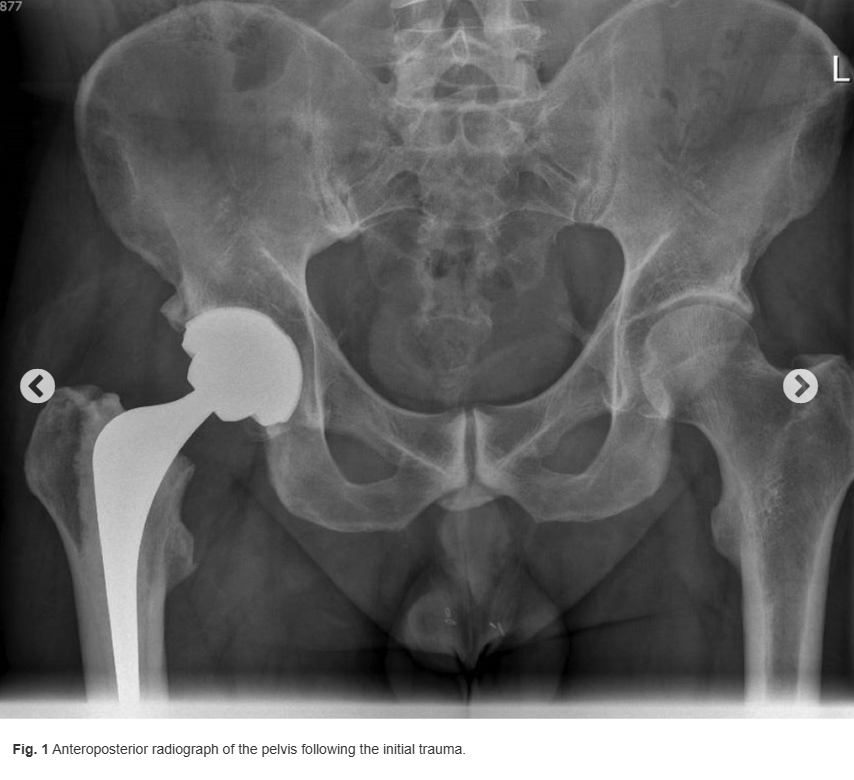
At 8 months following the primary surgical procedure, the patient slipped on black ice and fell heavily onto the right hip (trochanteric region). He was able to mobilize immediately afterward while experiencing only minimal pain. He was rereviewed by the operating orthopaedic surgeon within 1 week, and radiographs were taken, which were unremarkable (Fig. 1). The patient returned to normal activities soon after as he experienced pain relief. However, 1 month later, he noticed a sudden sharp pain and a sensation of grinding in the right hip following swimming. He was unable to bear weight and presented to the emergency department.
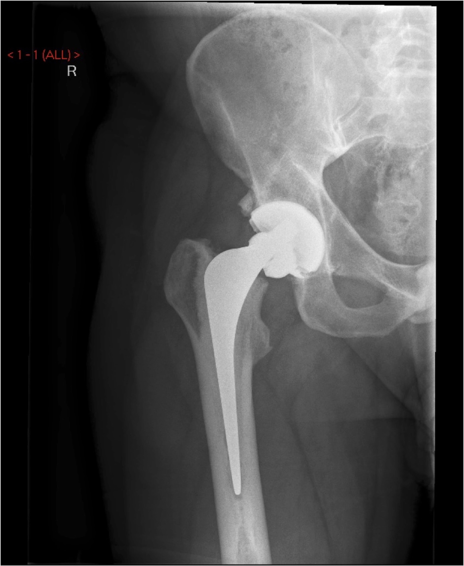
On examination in the emergency department, the patient had a well-healed posterior approach scar. There was no evidence of infection. He had tenderness to palpation of the groin. Passive and active ranges of movement were unrestricted, and the patient had some pain at 90° of passive hip flexion. He did not experience pain with internal and external rotations, but the examiner noticed palpable crepitus. Blood levels, including erythrocyte sedimentation rate (ESR) and C-reactive protein (CRP), were within normal limits. A radiograph of the hip is shown in Figure 2.
Related Suggestion
A 60-Year-Old Man with a Mass Following Hip Surgery
Clinical
A 60-Year-Old Man with a Mass Following Hip Surgery
1,647
2
Infrapatellar Fat Pad Modulates Osteoarthritis-Associated Cytokine and MMP Expression in Human Articular Chondrocytes
Knowledge
Osteoarthritis (OA) most frequently affects the knee joint and is associated with an elevated expression of cytokines and extracellular cartilage matrix (ECM), degrading enzymes such as matrix metalloproteinases (MMPs). Differences in gene expression of the intra-articularly located infrapatellar fat pad (IPFP) and other fatty tissue suggest its autonomous function, yet its role in OA pathogenesis remains unknown. Human IPFPs and articular cartilage were collected from OA patients undergoing total knee arthroplasty, and biopsies from the IPFP of healthy patients harvested during knee arthroscopy served as controls (CO). Isolated chondrocytes were co-cultured with either osteoarthritic (OA) or CO-IPFPs in a transwell system. Chondrocyte expression of MMP1, -3, -13, type 1 and 2 collagens, interleukin IL1β, IL6, IL10, and tumor necrosis factor TNFα was analyzed by RTD-PCR at day 0 and day 2, and TNFα secretion was analyzed by ELISA. The cytokine release in IPFPs was assessed by an array. Results: Both IPFPs (CO, OA) significantly reduced the expression of type 2 collagen and TNFα in chondrocytes. On the other hand, only CO-IPFP suppressed the expression of type 1 collagen and significantly induced the MMP13 expression. On the contrary, IL1β and IL6 were significantly induced when exposed to OA-IPFP. Conclusions: The partial loss of the suppressive effect on type 1 collagen gene expression found for OA-IPFP shows the pathological remodeling and dedifferentiation potential of the OA-IPFP on the chondrocytes. However, the significant suppression of TNFα implies that the OA- and CO-IPFP could also exhibit a protective role in the knee joint, preventing the progress of inflammation.
56
0
Comments 0
Please to post a comment~
Loading...
Likes
Send-Pen
Favorites
Comment