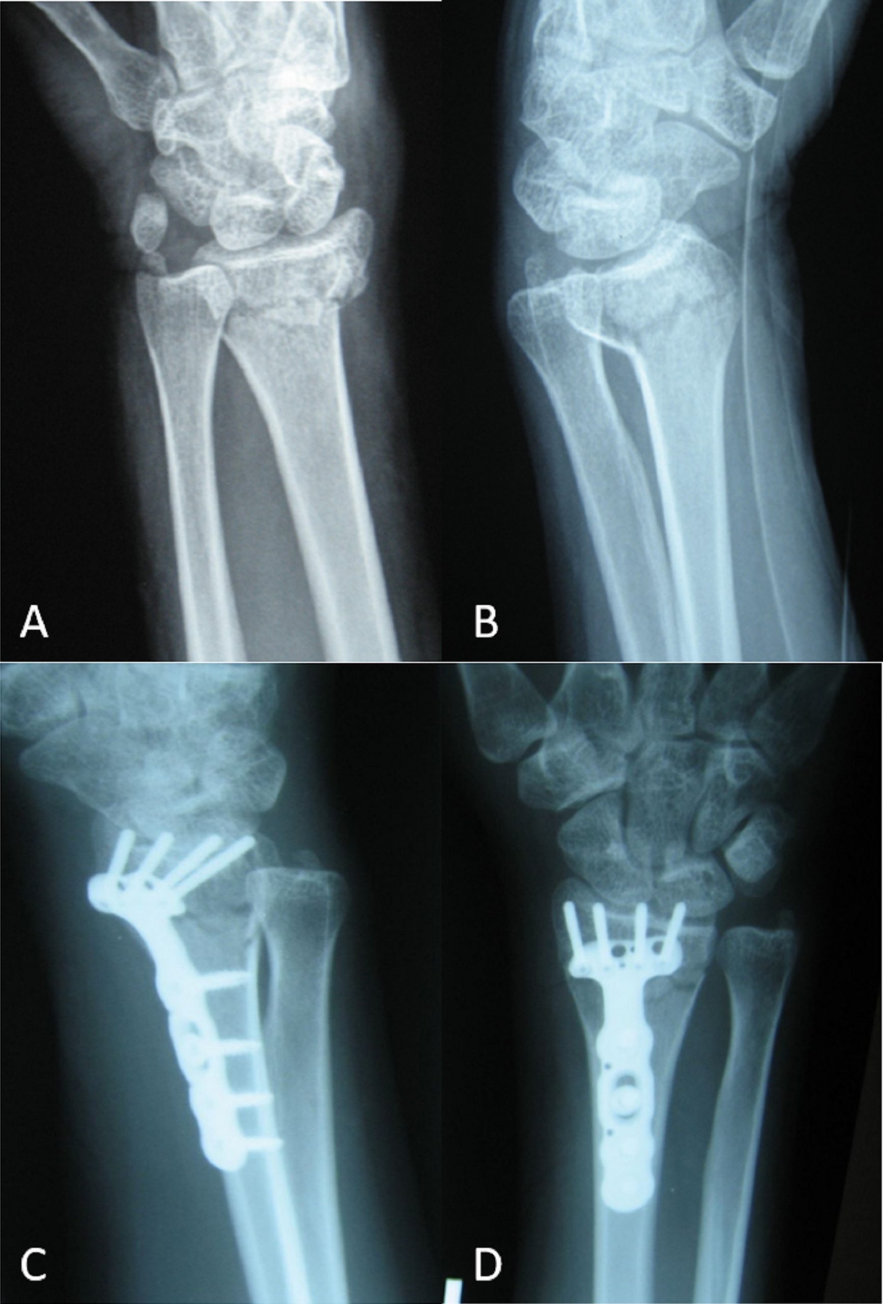
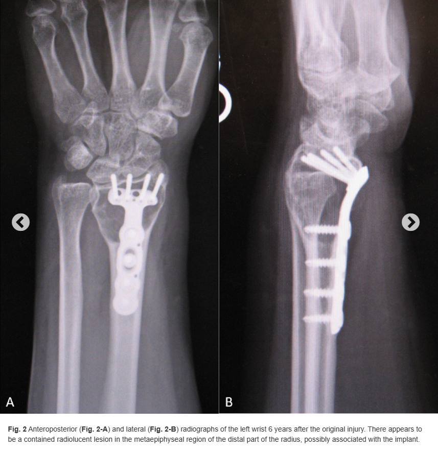
A 32-year-old woman experienced a distal radial fracture of the left wrist after trauma 6 years before presentation. At that time, she was found to have a displaced, extra-articular distal radial fracture without any evidence of an underlying lesion (Figs. 1-A and 1-B). She was treated with open reduction and internal fixation (ORIF) through a standard volar approach. There were no complications from the surgical procedure, and the postoperative course was unremarkable. Approximately 6 years later, when the patient was then about 10 weeks pregnant, she fell at her home after having had pain relief up until this event. After the fall, she presented with left wrist pain similar to when she initially fractured the wrist. No fracture was noted on radiographic evaluation, but a lesion was noted to have developed, possibly associated with the implant (Figs. 2-A and 2-B).


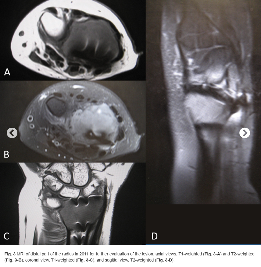
Magnetic resonance imaging (MRI) of the left distal part of the radius revealed extensive bulging of the cortex dorsally but no definite soft-tissue mass (Figs. 3-A through 3-D). The plan was to perform a procedure to remove the implant, obtain a frozen section to rule out malignancy, and perform intralesional curettage and fill the defect with bone cement.

Due to her pregnancy, obstetrics was consulted, and the patient received an axillary block for the procedure. The previous incision was used on the volar distal part of the radius. The plate was removed, and a frozen section was obtained. Of note, one screw was broken and the remaining piece was not able to be removed easily. Histology from the frozen section and the subsequently curetted lesion is shown in Figures 4-A and 4-B.

What is the diagnosis?

