


A 59-year-old man was referred by his family doctor with a history of a painful mass on the anteromedial aspect of the left thigh. The patient noticed that the mass had doubled in size over the course of 4 months. He reported no limitations in activity and there was no history of antecedent trauma to the area. The patient reported that he had not had any constitutional symptoms. He had no important medical or relevant family history.
Physical examination of the left thigh revealed a hard and mobile mass that was tender on palpation of the anteromedial aspect of the left femur distally. No associated signs of infection were observed. Neurologically, there were no motor or sensory deficits. Distal pulses were palpable, and the patient’s ipsilateral knee demonstrated a full range of motion. We did not observe lymphadenopathy or other masses.
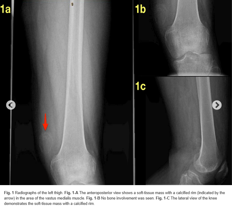
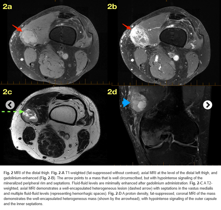
Radiographs of the left femur demonstrated a circumscribed 3.3 × 3 cm mass with a peripheral rim mineralization within the area of the vastus medialis muscle (Fig. 1). Magnetic resonance imaging (MRI) revealed a well-encapsulated, heterogeneous mass with high-intensity internal septations on T2-weighted imaging (Fig. 2). The lesion was bordered by a low signal intensity mineralized rim on T1-weighted imaging. There appeared to be fluid-fluid levels (Fig. 2-C). There was no bone involvement of the underlying normal bony structures.
Given the history of progressive and rapid enlargement of the mass, we proceeded with a core needle biopsy to rule out soft-tissue sarcoma.
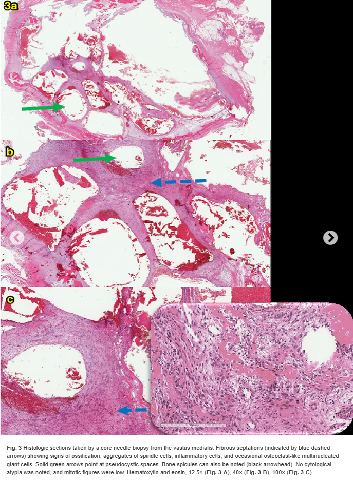
The needle biopsy specimen was interpreted as benign, so a decision was made to perform a complete excision of the soft-tissue mass because of the pain that the patient was experiencing. Surgical dissection showed a well-encapsulated mass, with no extension or attachment to the surrounding (normal) structure; resection of the entire tissue was performed. The resected specimen measured 4.5 × 4.5 × 2.5 cm and it contained red-brown, hemorrhagic soft-tissue fragments. The histology of the lesion is shown in Figure 3.
What is the diagnosis?
