


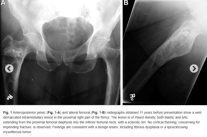
A 78-year-old man with multiple cardiovascular comorbidities presented to a musculoskeletal oncology clinic for acute right hip pain with a known right proximal femoral lesion. In 2006, he underwent lumbar and pelvic radiographs at our institution for chronic low back pain. The radiographs showed a radiolucent lesion in the right proximal part of the femur with a sclerotic rim and areas of internal septation and calcification (Figs. 1-A and 1-B). The lesion was believed to represent fibrous dysplasia or a liposclerosing myxofibrous tumor, and he was monitored with serial radiographs until he was lost to follow-up.
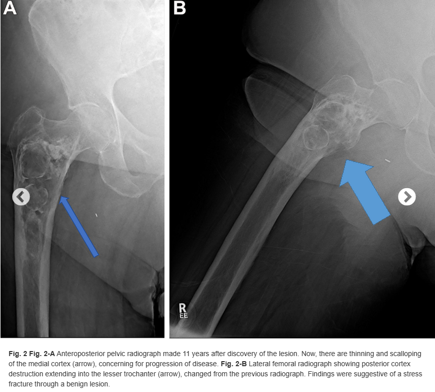
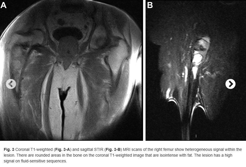
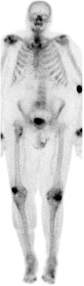
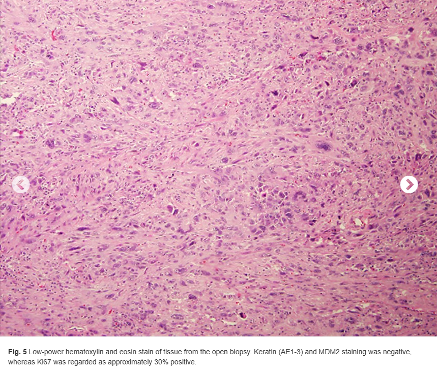
Eleven years later, the patient presented with a 2-month history of right proximal anterior thigh pain without inciting trauma. Radiographs of the right hip showed the proximal femoral lesion (Fig. 2-A). The lateral view (Fig. 2-B) showed loss of the posterior cortex at the level of the inferior lesser trochanter, representing a change in appearance since 11 years earlier. Magnetic resonance imaging (MRI) was obtained using a 0.2-T open magnet. (The MRI was of poor quality and could not be repeated because of insurance restrictions.) An intramedullary marrow-replacing lesion involving the proximal part of the femur and the heterogeneous signal was observed. The coronal T1-weighted image (Fig. 3-A) showed areas of high signal, isointense to subcutaneous fat. Fluid-sensitive sequences showed areas of high signal throughout the lesion, although fat-containing areas were hypointense, as expected. Linear areas of low signal consistent with mineralization were appreciated, corresponding to the radiographic appearance. On the sagittal short tau inversion recovery (STIR) image (Fig. 3-B), posterior perilesional soft-tissue extension was identified. A bone scan showed mild radiotracer uptake in the proximal part of the femur, with an area of moderately increased uptake along the medial proximal femoral cortex (Fig. 4). Histology from an open biopsy is shown in Figure 5.

What is the diagnosis?